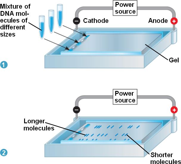
The longest molecules migrate slower than the shorter molecules in gel electrophoresis.
Gel electrophoresis is
a technique used to separate DNA molecules. DNA molecules of various
sizes are first placed in a tray filled with a gel at the negatively
charged cathode. The positively charged anode is located at the
opposite end of the tray. Since DNA is negatively charged, all DNA
molecules will move towards the anode because its positive charge
attracts the DNA. Now, imagine a river into which rocks of various sizes
have been dropped: the largest rocks will move the least, while the
smallest rocks will move the most. Likewise, the longest DNA molecules
move the least, and the shortest DNA molecules move the most because the
gel restricts movement. DNA molecules of each specific size will form a
band, and each different band corresponds to a DNA molecule of a
different size, with the smallest molecules closest to the anode. (8)
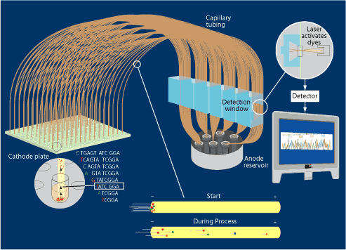
In capillary electrophoresis, molecules separate out by size as they move from the negatively charged cathode to the positively charged anode. A laser causes the dye on the last nucleotide to fluoresce, and a computerized detector records the information.
Capillary electrophoresis is
a type of gel electrophoresis that uses a thin tube called a capillary
in place of a tray. Also, a very sensitive type of gel called acrylamide
gel that can separate DNA molecules have only a one base-pair
difference in length and the DNA molecules are placed in an electrolyte
solution. As
DNA molecules migrate towards the positively charged anode and separate by size, a detector can detect the
length of each DNA molecule and the fluorescent nucleotide at the end of the molecule. (The fluorescent nucleotide flashes a certain color when exposed to the laser. Each of the four nucleotides is assigned a different color, as shown below.) (52)
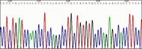 | On the left is a sample detector output. Each nucleotide is represented by a different color dye. |